Confocal
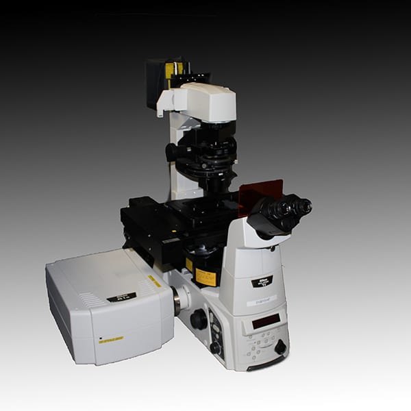
Nikon A1Rsi
The Nikon A1Rsi is a scanning confocal microscope is suitable for a broad range of applications including spectral and rapid imaging. The system is equipped with a hybrid scan head, incorporating both high-resolution galvo and an ultra-high speed resonant scanners, enabling simultaneous photo-stimulation and image acquisition. The scan head is equipped with two GaAsP PMTs, and two high-sensitivity PMT detectors as well as a transmitted light PMT for DIC imaging.
System configuration
- Laser lines: 405, 445, 488, 514, 561 and 640nm
- Objectives: 10X and 20X dry and 40X, 60X and 100X high NA oil immersion
- Motorized stage: xy-stage including a nano-positioning piezo z-insert
- Incubation: Tokai-hit stage-top incubator capable of temperature, humidity, CO2 and O2 regulation
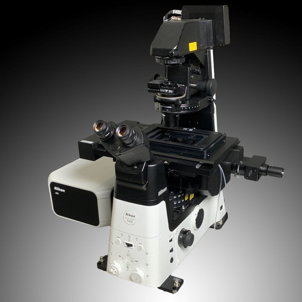
Nikon AX-R
The Nikon AXR is a laser scanning confocal microscope, designed for a broad range of applications, including live cell imaging, three-dimensional reconstructions, FRAP/FRET analysis, large area scanning, and spectral analyses. The system is based on the Ti2 microscope stand, which includes the Perfect Focus stabilization system and an automated water immersion dispenser, and features a 25mm wide field of view. The scanhead features an 8k galvo scanner as well as a 2k resonant scanner capable of rapid image acquisition and enabling simultaneous photobleaching/photostimulation and image acquisition. The detectors include 4 GAsP detectors for visible range detection, an alkali PMT for Cy7/AF 750 NIR detection, and a spectral detector for hyperspectral images in up to 66 emission channels for unmixing.
System configuration
- Laser lines: 405, 445, 488, 514, 561, 640, and 730 nm
- Objectives: 2X, 10X and 20X dry, 20X and 40X water, 25X silicone, 60X and 100X high NA oil immersion
- Motorized stage: xy-stage
- Prior nanopositioning piezo z-insert with 650µm travel range
- Incubation: Tokai-hit stage-top incubator capable of temperature, humidity and CO2 regulation
Olympus FluoView1200
The Olympus FV1200 scanning confocal microscope is a versatile imaging platform for both fixed and living samples. The system is equipped with two GaAsP PMTs and three high-sensitivity PMT detectors for five-color confocal imaging, and a transmitted light PMT for DIC imaging. The system is capable of acquiring spectral image data at 2nm resolution. The platform is also equipped with a high-speed SIM laser scanner enabling simultaneous photo-bleaching / photo-activation and image acquisition
System configuration
- Laser lines: 405, 440, 488, 515, 559 and 635nm
- Objectives: 10X dry, 10X, 20X and 40X water immersion, 20X (corr), 100X oil immersion and 60X super chromatic aberration corrected oil immersion
- Motorized stage: xy-stage
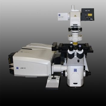
Zeiss LSM 880 Confocal with Airyscan
The Zeiss LSM 880 Airyscan confocal microscope is suitable for a broad range of applications including spectral and rapid imaging. The system is equipped with a unique scan head, incorporating a high-resolution galvo scanner along with two PMTs and a 32-element spectral detector as well as a transmitted light PMT for DIC imaging. In addition, the Airyscan unit provides sub-diffraction limited imaging down to 120nm resolution and the FAST beam shaper enables imaging at up to 27 fps.
System configuration
- Laser lines: 405, 458, 488, 514, 561 and 633nm
- Objectives: 10X and 20X dry and 40X, 60X and 100X high NA oil immersion
- Motorized stage: xy-stage including a nano-positioning piezo z-insert
- Incubation: Pecon stage-top incubator capable of temperature, humidity and CO2 regulation
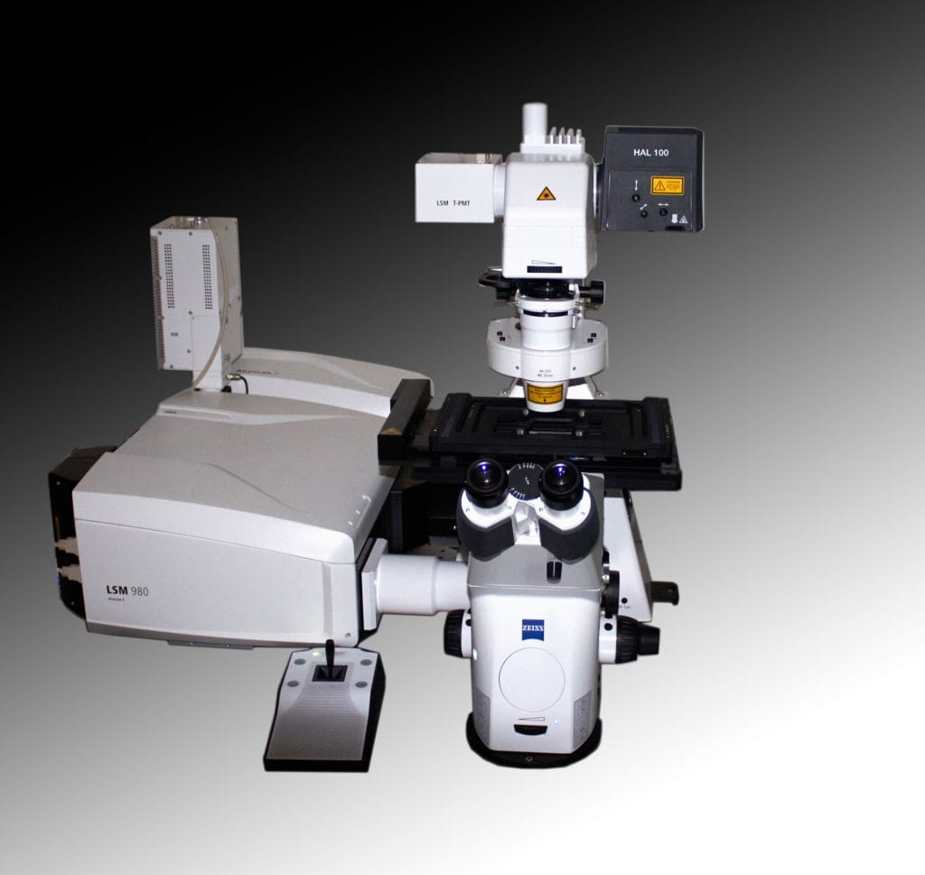
Zeiss LSM 980 with Airyscan 2
The Zeiss LSM 980 Airyscan 2 confocal microscope is suitable for a broad range of applications including spectral and rapid imaging. The system is based on a motorized inverted Zeiss Axio Observer Z1 microscope frame. The system is equipped with a unique scan head, incorporating a high-resolution galvo scanner along with two PMTs and a 32-element spectral detector as well as a transmitted light PMT for simultaneous DIC imaging.The system also includes a dedicated PMT for imaging NIR fluorophores such as AlexaFluor 750. Lastly, the Airscan 2 unit provides sub-diffraction limited imaging, down to 120nm in the lateral direction, as well as several parallelization modes for capturing spatial information at up to 9.6 Hz at full field of view.
System configuration
- Laser lines: 405, 458, 488, 514, 561, 639, and 730 nm
- Objectives: 10X and 20X dry, 40X water, and 63X high NA oil immersion
- Motorized stage: xy-stage
- Incubation: Pecon stage-top incubator capable of temperature, humidity and CO2 regulation
Live Cell Confocal
Nikon Spinning Disk Confocal
The Nikon Spinning Disk (SD) confocal microscope is designed for high-magnification 4D imaging of living cells with minimal effects of photo-damage. The system is fitted with a Yokagawa CSU-X1 variable speed Nipkow spinning disk scan head along with an Andor TuCam adapter that enables simultaneous two-color confocal imaging at video-rate (30fps) on two separate Andor Zyla sCMOS cameras. The system also boasts an LED-based DMD system for ultrafast photo-stimulation.
System configuration
- Laser lines: 405, 488, 561 and 640nm
- Objectives: 10X and 20X dry, 60X and 100X high NA oil immersion and 40X water immersion (corr).
- Motorized stage: xy-stage including a nano-positioning piezo z-insert
- Ti LAPP DMD (Deformable Mirror Device) LED source for ultrafast photo-stimulation
- Miniscanner galvo-based laser scanning system for FRAP / photo-activation
- Incubation: Tokai-hit stage-top incubator capable of temperature, humidity, CO2 and O2 regulation
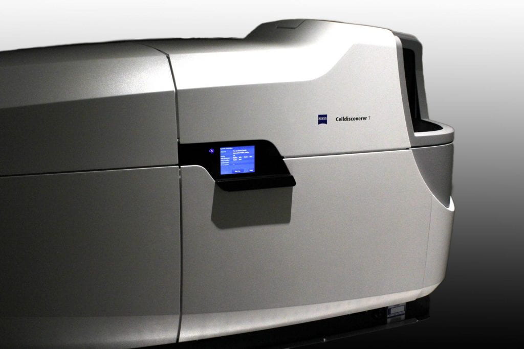
Zeiss Cell Discoverer 7
The Zeiss Celldiscoverer 7 is a fully automated plate reading live-cell microscope suitable for a broad range of applications including multi-color-fluorescence imaging capable of both widefield and confocal modalities. The system is based on inverted optics and is equipped with a Colibri 2 LED illumination source and high-sensitivity sCMNOS camera for widefiueld imaging and a LSM900 scan head for confocal imaging. The LSM900 scan head is equipped with 2 GaAsP PMTs for simultaneous two-color detection, or sequential, 4 color detection.
System configuration
- 2 x Colibri 2 LED illumination source
- Laser lines: 405, 488, 561 and 638nm
- Objectives: 5X and 20X for plastic bottoms dishes, 20X dry and 50X water immersion for glass bottom dishes
- Tube-lenses: 0.5X, 1X amd 2X tube lenses for additional magnification
- Motorized stage: xy-stage
- Incubation: Full incubation system capable of temperature, humidity and CO2 regulation
Widefield
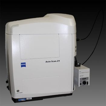
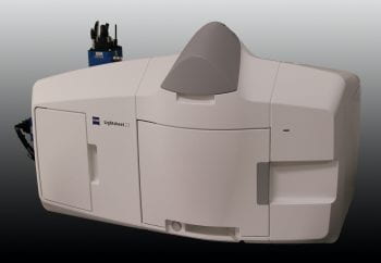
Zeiss AxioScan .Z1
The Zeiss AxioScan Z1 is a fully automated, 100-slide capacity, brightfield / fluorescence slide scanning system equipped with a mercury illumination source and high-speed filter wheel fitted with filters for DAPI, GFP, RFP and far-red fluorescence imaging. The system is equipped with two cameras; a Hammatsu Orca Flash sCMOS camera for high-speed fluorescence and a color CCD camera for histological imaging of H&E, peroxidase and DAB stained slides.
System configuration
- HXP120 excitation source
- Objectives: 5X, 10X, 20X and 40X dry
- High accuracy motorized stage
- 100-slide capacity loader
- Cassettes for 4 x 1×3 slides and 2 x 2×3 slides
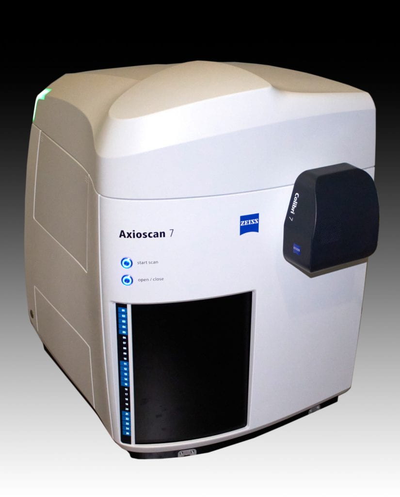
Zeiss AxioScan 7
The Zeiss AxioScan 7 is a fully automated, 100-slide capacity, brightfield / fluorescence slide scanning system equipped with a Colibri LED illumination source for rapid fluorescent imaging of blue, green, red, far red and near-IR fluorescent probes. The system is equipped with two cameras; a Hammatsu Orca Flash sCMOS camera for high-speed fluorescence and a color CCD camera for histological imaging of H&E, peroxidase and DAB stained slides.
System configuration
- Colibri 7 LED illumination sources
- Objectives: 5X, 10X and 20X dry
- High accuracy motorized stage
- 100-slide capacity loader
- Cassettes for 4 x 1×3 slides and 2 x 2×3 slides
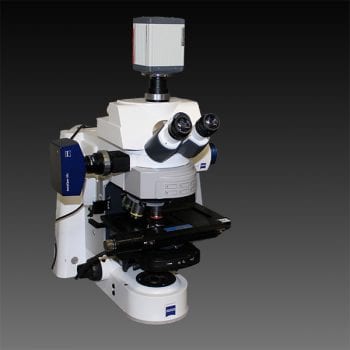
Zeiss AxioImager Z2
The Zeiss Axio Imager Z2 is a motorized upright brightfield / fluorescence microscope equipped with an ApoTome 2 optical sectioning grid imager. The system is equipped with an LED illumination source and filtersets for DAPI, GFP, RFP and far-red imaging as well with the relevant prisms and sliders for Differential Interference Contrast Microscopy (DIC). The system is equipped with two cameras; a Hammatsu Orca Flash sCMOS camera for fluorescence and a color CCD camera for histological imaging.
System configuration
- Excite LED excitation source
- Objectives: 5X, 10X, 20X dry and 60X and 100X oil and 40X water immersion (corr) lenses
- Motorized stage: xy-stage
Zeiss AxioZoom V16
The Zeiss AxioZoom V16 is a motorized fluorescent and brightfield macroscope, equipped with a Plan Neofluoar Z 1x/0.25 objective, with a 7.0-112x continuously variable zoom range. The system has filter sets for DAPI, GFP, and RFP, as well as brightfield diascopic and episcopoc illumination. Acquisition is performed on either a Hamamatsu Orca Flash sCMOS for high-speed fluorescence, or a Zeiss AxioCam HRC color camera for histological stained samples.
System configuration
- HXP120 excitation source
- Objective: Plan Neofluoar Z 1x/0.25 (7x-112x zoom)
- Motorized stage
Two-Photon
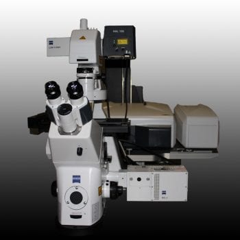
Zeiss 880 Two-Photon (inverted)
The Zeiss LSM 880 Airyscan two-photon microscope is suitable for a broad range of applications in scattering media. The system is equipped with a unique scan head, incorporating a high-resolution galvo scanner along with two PMTs and a 32-element spectral detector as well as a transmitted light PMT for DIC imaging. In addition, the Airyscan unit provides sub-diffraction limited imaging down to 120nm resolution and the FAST beam shaper enables imaging at up to 27 fps.The system is also equipped with two non-descanned detectors (NDDs) for two-photon imaging in thick tissue samples.
System configuration
- Laser lines: 405, 458, 488, 514, 561 and 633nm and a Coherent Discovery laser with a tunable 680-1300nm line and a static 1040nm line
- Objectives: 10X and 20X dry and 40X, 60X and 100X high NA oil immersion
- Motorized stage: xy-stage including a nano-positioning piezo z-insert
- Incubation: Pecon stage-top incubator capable of temperature, humidity and CO2 regulation
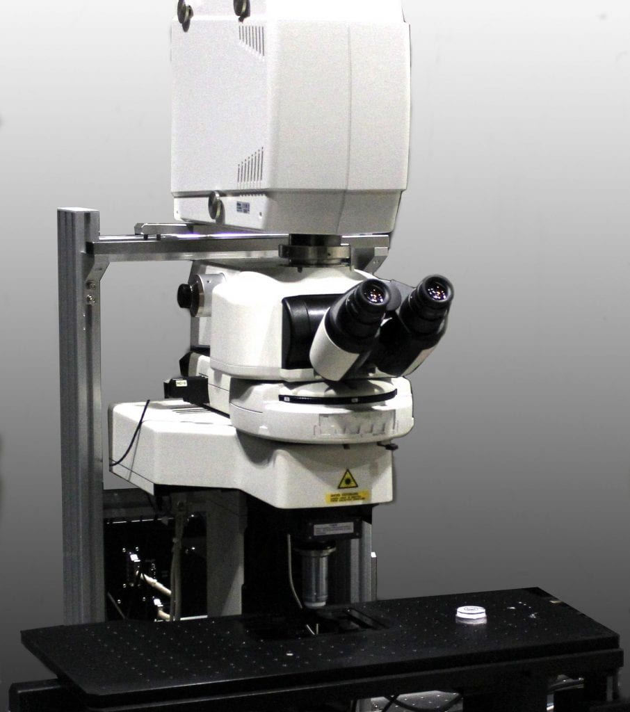
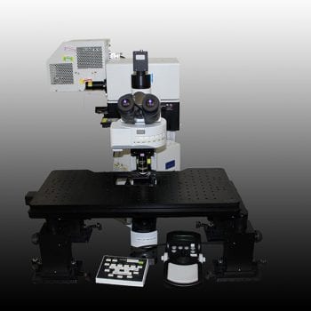
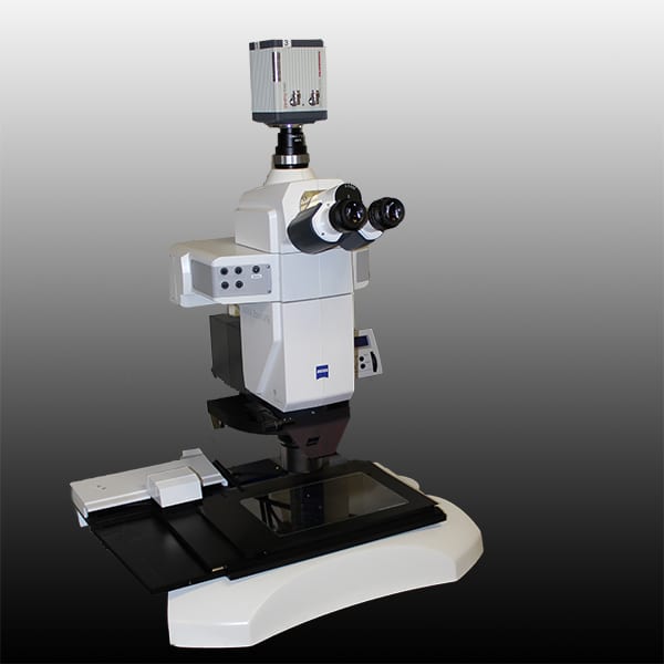
Nikon A1RHD25 MP Two-Photon (upright)
The Nikon A1RHD25 MP multi-photon microscope is suitable for a broad range of applications including spectral and rapid imaging as well as deep tissue imaging. The system is based on a motorized Nikon FN1 upright microscope frame. The system is equipped with a unique scan head, incorporating a high-resolution 8kHz resonant galvo scanner along with four PMTs (2 GaAsP) detectors as well as a transmitted light PMT for simultaneous DIC imaging. The system is also equipped with four non-descanned detectors (NDDs) for two-photon imaging in thick tissue samples and live animals
System configuration
- Laser lines: 405, 488, 561 and 633nm and tunable Titanium Sapphire femtosecond infrared laser
- Objectives: 10X and 20X dry, 16X and 25X water dipping, and 10X RI-Matching multi-immersion
- Motorized stage: xy-stage
Super-resolution
Nikon n-SIM Structured Illumination
The Nikon n-SIM microscope allows for sub-diffraction super-resolution imaging using high-frequency structured illumination in three-dimensions for resolutions at 100nm. The system is equipped with two Andor iXon 3 EM-CCD cameras fitted to an Andor TuCam dual camera adapter, which facilitates simultaneous two-color and sequential four-color super-resolution imaging using either 2D-TIRF SIM or 3D SIM imaging modes.
System configuration
- Laser lines: 405, 488, 561 and 640nm
- Objectives: 60X water immersion and 100X oil immersion high NA lenses
- Motorized stage: xy-stage including a nano-positioning piezo z-insert
- Incubation: Tokai-hit stage-top incubator capable of temperature, humidity and CO2 regulation
Nikon n-STORM/TIRF
The Nikon n-STORM TIRF Microscope allows imaging close to the surface of a coverslip facilitating imaging of endo- and exocytic processes as well membrane trafficking events. The system is equipped with both an Andor Zyla 4.2 Megapixel sCMOS camera when imaging in TIRF mode or an Andor iXon 3 EM-CCD when imaging in single-molecule or STORM (STochastic Optical Reconstruction Microscopy) super-resolution imaging mode.
System configuration
- Laser lines: 405, 488, 561 and 640nm
- Objectives: 10X and 20X dry and 60X and 100X oil immersion high NA lenses
- Motorized stage: xy-stage including a nano-positioning piezo z-insert
- Incubation: Tokai-hit stage-top incubator capable of temperature, humidity and CO2 regulation
Lightsheet
Zeiss Lightsheet 7
The Zeiss Lightsheet 7 planar illumination microscope is suitable for imaging dynamic processes in developing organisms, live organoid cultures as well as the anatomical content of cleared tissues. The system is configured with dual lightsheet illumination arms and a unique pivot scan mechanism to avoid striping artifacts. In addition, the system is equipped with a live-cell incubation system enabling the physiological imaging of living specimens.
System configuration
- Laser lines: 405, 488, 561, and 638 nm
- Objectives: 2.5X and 5Xfoc dry and 10X, 20X, 40X and 63X water immersion and 20X (1.38ri – CLARITY), 20X (1.45ri – SCALE), and 20X (1.53-DISCO) (Note +/- 0.03 on RI)
- Dual Lightsheet illumination with Pivot Scanning Technology and foc optics
- Dual PCO.Edge CMOS Cameras for simultaneous two-color acquisition
- Adjustable mesochamber for whole-organ imaging
- Motorized stage: xyz-stage
- Incubation: System capable of temperature, humidity and CO2 regulation
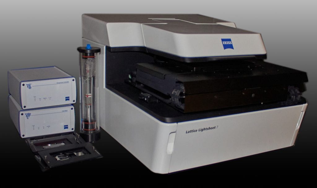
Zeiss Lattice Lightsheet 7
The Zeiss Lattice Lightsheet 7 microscope is suitable for imaging highly dynamic processes in living cells and tissues as well as live organoid cultures. The system is configured with multiple lattice options yielding different lightsheet thicknesses and axial resolutions In addition, the system is equipped with a iBidi live-cell incubation system enabling the physiological imaging of living specimens.
System configuration
- Laser lines: 488, 561 and 638nm
- Objective: 44x 1.0NA
- Camera: PCO.edge sCMOS camera
- Lattice sheet thicknesses: 550nm, 700nm 1400nm
- Motorized stage: xyz-stage
- Incubation: System capable of temperature, humidity and CO2 regulation
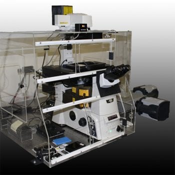
OCPI-II
OCPI-II is a home-built Light-Sheet Fluorescence Microscope (LSFM) based on an Objective-Coupled Planar Illumination (OCPI) implementation. This system excels at high-speed volumetric imaging of water-immersed fluorescent samples. This approach it is well suited to analyzing large ensembles of neuronal elements or for tracking cellular calcium. The microscope is equipped with two high speed cameras that allow dual-color imaging. A piezoelectric positioner allows rapid z-scanning up to 800um of travel.
System configuration
- Laser lines: 405, 440, 488, 515 and 561nm
- Objectives: 10X/0.3 NA, 20X/0.5 NA, 4X/0.28 NA , 10x/0.6 NA
- A 2-sCMOS camera system for the simultaneous imaging of two colors, including ratiometric imaging
- 8 Configurable TTL outputs for synchronizing custom equipment / triggering stimuli
- A multi-channel perfusion system with temperature control for maintaining physiological conditions
- Micro-manipulators for simultaneous electrophysiology and imaging
- An open, upright stage configuration allowing introduction of addifional components, such as inlets, for pharmacological agents, stimulating electrodes and a treadmill for in vivo mouse experiments
- Imaging speed for a 500um cubic volume sampled with 5 um z-spacing: ~2 volumes per second (exact value depends on objective, forthcoming upgrades will increase this further)