Cryo-Transmission Electron Microscopy
Titan Krios G3 300kV Cryo-TEM
The FEI Titan Krios G3 300kV Cryo-TEM is the premier system for both single particle averaging and cryo-electron tomography of vitrified biological sThe FEI Titan Krios G3 300kV Cryo-TEM is the premier system for both single particle averaging and cryo-electron tomography of vitrified biological samples. The system is equipped with an image corrector, an auto-loading system, and two detection cameras: a CMOS (Ceta) and an energy-filtered Direct Detector (Thermo Fisher Falcon 4i and Selectris X Energy Filter) for low dose imaging. amples. The system is equipped with an image corrector, an auto-loading system, two detection cameras, a CMOS (Ceta) and an energy-filtered Direct Detector (Gatan K2 Summit on GIF filter) for low dose imaging. In addition, the system is also equipped with a phase plate and STEM detector.
System configuration:
- Sample Auto-loading System – allows twelve independent samples to be loaded into a cassette, which can be transferred into the auto-loading system for delivery into the Titan Krios for imaging. The auto-loader is maintained at liquid nitrogen temperatures to protect the samples from any environmental factors, and each individual sample holder (AutoGrid) is robust and protects the sample against deformation and / or damage.
- Ceta CMOS camera – The Ceta is a 16 Megapixel digital camera designed specifically for regular TEM/STEM imaging. The large field of view and small pixel size (a characteristic of CMOS sensors) allows a fast read-out for dynamic experiments and is capable of recording full-frame 4k x 4k images at 30 fps and a reduced field of 512 x 512 at > 250 fps. The camera can also easily switch between this fast mode and high-resolution mode with ease.
- Thermo Fisher Falcon 4i and Selectris X Energy Filter – The combined Falcon 4i direct electron detector and Selectris X energy filter allows for the direct detection of electrons at high sensitivity. In addition, the presence of the energy filter allows the preferential detection of zero-loss electrons, thus serving as an additional mechanism to enhance the contrast of unstained, vitrified biological specimens. This new addition increases the signal-to-noise ratio significantly for both single particle and tomography acquisitions.
- Cs Aberration Corrector – The C-TWIN objective lens with its larger pole-piece gap is essential for high tilt angle tomographic acquisitions. Its symmetric design allows for the changing of the accelerating voltage without adjusting the stage z-position. Additionally, it ensures that there is no performance compromise, or any re-alignment between TEM and STEM imaging modes. However, whilst important for tomography, the larger pole-piece gap in the C-TWIN objective lens increases spherical aberration. The addition of an image corrector removes this added spherical aberration (Cs), thus restoring the TEM/STEM resolution to that of a Titan microscope platform with a smaller pole-piece separation.
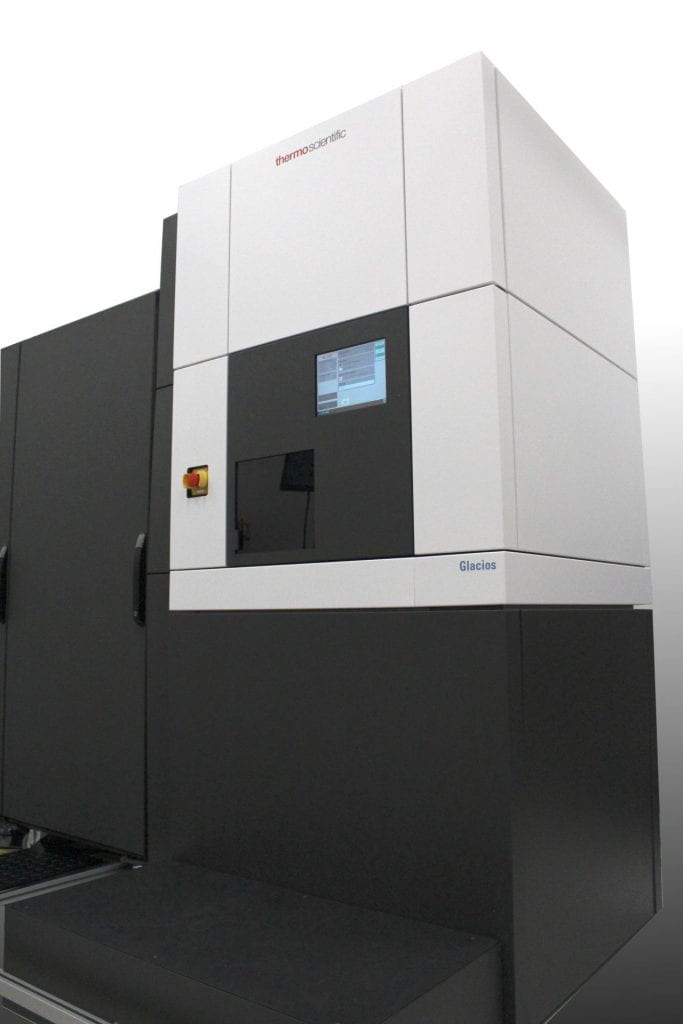
Glacios 200kV Cryo-TEM
The ThermoFisher Scientific Glacios 200kV Cryo-TEM is the premier mid-range platform for the near-atomic resolution imaging of vitrified proteins and macromolecular assemblies. The system is equipped with an auto-loading system, three detection cameras, a CMOS camera optimized for diffraction applications (Ceta-D), and two direct electron detection devices, a Falcon IV and Gatan K3 for low dose imaging. In addition, the system is also equipped with a phase plate and STEM detector.
System configuration:
- Sample Auto-loading System – allows twelve independent samples to be loaded into a cassette, which can be transferred into the auto-loading system for delivery into the Titan Krios for imaging. The auto-loader is maintained at liquid nitrogen temperatures to protect the samples from any environmental factors, and each individual sample holder (AutoGrid) is robust and protects the sample against deformation and / or damage.
- Ceta-D CMOS camera – The Ceta is a 16 Megapixel digital camera designed specifically for regular TEM/STEM imaging and is optimized for diffraction applications such as micro-ED. The large field of view and small pixel size (a characteristic of CMOS sensors) allows a fast read-out for dynamic experiments and is capable, of recording full-frame 4k x 4k images at 30 fps and a reduced field of 512 x 512 at > 250 fps. The camera can also easily switch between this fast mode and high-resolution mode with ease.
- Falcon IV Direct Electron Detector – The Falcon IV allows for the direct detection of electrons at high sensitivity. This 4th generation DED from ThermoFisher Scientific has the highest DQE over the entire spatial frequency range of any DED.
- Gatan K3 Direct Electron Detector – The Gatan K3 allows for the direct detection of electrons at high sensitivity. This new platform, redesigned from the ground-up is a 24 megapixel sensor, capable at recording at 1,500 fps, almost 4 times faster than the previous K2 camera.
- Volta Phase Contrast System – A major issue in the imaging of unstained, vitrified samples is the lack of electron density, and thus contrast in the acquired images. A new, and cutting-edge approach that almost entirely removes the need for the addition of beam defocus is phase contrast imaging. By separating the phases of elastically and inelastically scattered electrons that pass through the sample by 90o, the in-focus contrast in the imaging plane can be drastically improved, which is beneficial in the imaging of weak phase objects, such as vitrified biological specimens.
- STEM detector – The ability to scan the electron beam whilst acquiring the transmitted electron signal, a so-called “STEM” image is essential in high-resolution studies of cellular macromolecules. The incorporation of STEM functionality onto the Glacios platform increases its versatility without compromising any of the other imaging modes of the microscope (TEM, EFTEM etc).
Cryo-Focused Ion Beam Microscopy
Aquilos 2 Cryo-FIB
The Thermo Scientific Aquilos 2 Cryo-Focused Ion Beam Microscope is an advanced platform for the preparation of thin lamellae of vitrified cells and bulk-frozen tissues for cryo-Electron Tomography workflows. The Aquilos 2 is equipped with a NICol UHR non-immersion SEM column containing a Schottky field emission gun optimized for high brightness/high current facilitating the low-noise imaging of frozen biological samples at a resolution of 6 nm at 2kV. The Aquilos 2 is also configured with multiple detection options including:
- Everhart-Thornley Secondary Electron Detector – optimized for use across the available kV and current range
- In lens detection is comprised of a segmented, lower in-lens detector (T1) and an upper in-lens detector (T2)
- ICE detector – for secondary ion and electron detection
- Integrated infrared-CCD camera – for in-chamber viewing of the sample
- Nav-Cam color optical camera – for taking top-down images of samples for navigation purposes
FIB Source:
The Aquilos 2 is equipped with a Sidewinder Ga-Liquid Metal Ion Source (Ga-LMIS) column that is capable of generating a focused beam of gallium ions that has a precision milling resolution of 7 nm at 30kV.