The WUCCI provides a complete service pipeline for light, electron, ion and 3D X-ray microscopy which is comprised of the following elements:
- Affordable access to state-of-the-art light, electron, ion and 3D X-ray microscopy
- Expert support in assay design, training on instrumentation and assistance with data acquisition
- Analysis of multi-dimensional imaging datasets using commercial or custom-written algorithms
Service Requests

Tissue Clearing Sample Preparation
SEM Sample Preparation
TEM Sample Preparation
FIB-SEM Sample Preparation
Deep Etch EM Sample Preparation
XRM Sample Preparation

Other Services
Cryo-EM Project Request
Project Request Form
Training Requests
Request Training
Imaging types available in the WUCCI
- Widefield Microscopy: Multi-color fluorescence and DIC imaging of fixed samples.
- Confocal Microscopy: Three-dimensional multi-color fluorescence imaging of fixed mounted cell cultures or tissue sections as well as live cell cultures and living specimens such as C. elegans and zebrafish model systems.
- Super-Resolution Microscopy: 2D / 3D localization microscopy, or Structured Illumination Microscopy of fixed cell cultures or thin tissue sections.
- Two-Photon Microscopy: Three-dimensional multi-color fluorescence imaging of fixed mounted thick tissue sections as well as live specimens such as in vivo mouse imaging.
- Transmission EM: Ultrastructural nanoscale imaging of cell cultures and tissue samples as well as purified proteins / complexes.
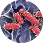
- Scanning EM: Ultrastructural topographic imaging of cell cultures and tissue samples.
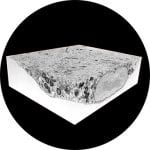
- Focussed Ion Beam-SEM: Three-dimensional serial-block face (SBF) nanotomography of cell cultures and tissue samples.
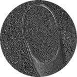
- Non-invasive sub-micron-level resolution tomographic imaging of hard and soft materials from microns to many centimeters in size.
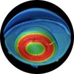
- Single Particle Cryo-EM: Atomic-level resolution reconstruction of macromolecular structures.
- Cryo-Electron Tomography: Three-dimensional reconstruction of viral particles and thinned lamella of vitrified cell cultures.