Dear Colleagues, The Washington University Center for Cellular Imaging (WUCCI) is delighted to announce that following an international search, Eduardo Rosa-Molinar, Ph.D. has accepted our offer to become the new scientific director of the WUCCI. Dr. Rosa-Molinar will also be appointed Professor of Cell Biology & Physiology and Neuroscience. Dr. Rosa-Molinar has an extensive record of impactful […]
Author: peterbayguinov
WashU Hosts Cryo-EM Symposium
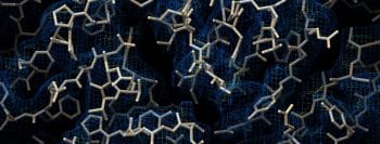
December 7, 2023 | 1:00pm – 4:00pm Join us at Washington University in St. Louis to learn about the adoption of Cryo Electron Microscopy (cryo- EM) as a versatile, multidisciplinary approach to analyze protein structure. Using cryo-EM, scientists can directly image and analyze structural details in 3D, enabling them to characterize mechanisms of action, discover […]
Fitzpatrick Awarded NIMH S10 Instrument Grant
Six investigators from the Departments of Genetics and Neuroscience at the School of Medicine will make use of this imaging platform to enable a wide-range of studies aimed at investigating the genetic regulation of synapse-localized translation, the molecular mechanisms of gene regulation as well as the discovery and characterization of genetic etiological factors involved in Autism Spectrum Disorder (ASD), how mGluR5 mediated signal transduction is implicated in anxiety and depression and how loss of hippocampal activity leads to learning and memory deficits in cognitive disease.
Fitzpatrick Awarded Chan Zuckerberg Initiative Imaging Scientist Fellowship
Dr. Fitzpatrick will work to innovate novel approaches and resources for training and educating users and will facilitate outreach efforts in order to train the next generation of imaging scientists.
WUCCI Welcomes Sanja Sviben
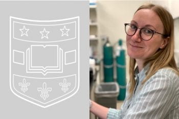
Dr. Sviben received her degree from the Max Plank Institute where she did some phenomenal work using Scanning Electron Microscopy to study biomineralization.
RBC Fidelity Paper Out with Doctor Lab
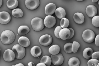
Red blood cell phenotype fidelity following glycerol cryopreservation optimized for research purposes
Compound C Paper Out with You Lab
Compound C inhibits nonsense-mediated RNA decay independently of AMPK
NINDS Awards U01 for A-syn Cryo-EM
In this project, we will use cryo-electron microscopy (cryo-EM) to determine atomic resolution structures of Asyn fibrils in Lewy body dementia, in conjunction with solid-state NMR (SSNMR) for refinement of structures.
Fitzpatrick Awarded NIH HEI S10 Grant
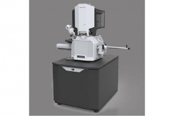
This microscope system will allow innovative nanomanipulation of vitrified cells and tissues into thin lamellae (~300 nm thickness) for Cryo-Electron Tomography (Cryo-ET), and will facilitate the in situ elucidation of three-dimensional structures of cellular organelles and macromolecular complexes at nanometer resolution.
PLAGL2 Paper Out with the Madison Lab
The Zinc Finger Transcription Factor PLAGL2 Enhances Stem Cell Fate and Activates Expression of ASCL2 in Intestinal Epithelial Cells
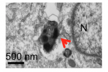
3D EM of BMA with Scheller/Craft Labs
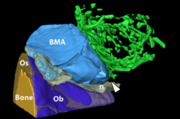
Characterization of the bone marrow adipocyte niche with three-dimensional electron microscopy
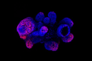
WUSTL Movie Wins BioArt Competition

A video of a lacy network of red, blue, gold and green strands, tracing the many blood vessels in a mouse’s lungs, was named a winner of the 2017 BioArt competition.